ESC
Computational & Automation

Coding, Automation & Data Science

I build computational pipelines, automate lab workflows, and integrate advanced microscopy with quantitative analysis to generate reproducible, decision-enabling datasets at scale.

Python AutoMorphoTrack BRAVO / Tecan / Hamilton 96/384-Well Miniaturization Machine Learning Morphology Classification Organelle Tracking Open Source
01

AutoMorphoTrack Software

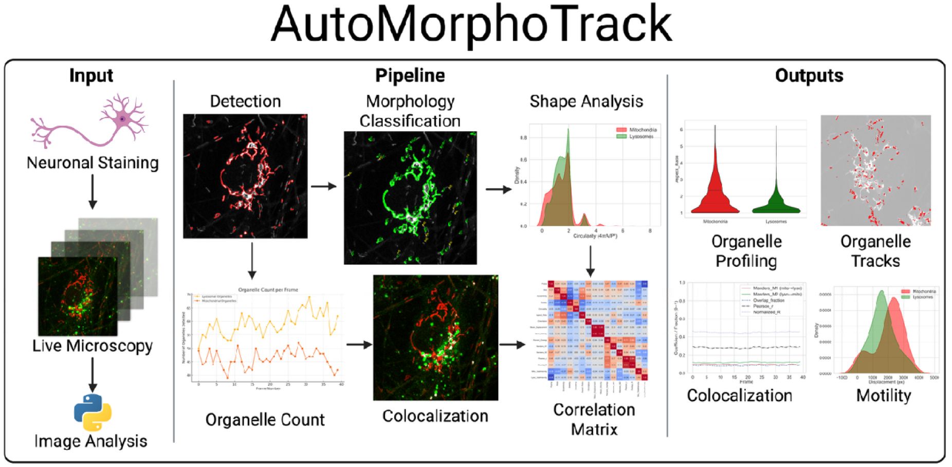
AutoMorphoTrack is my open-source Python package for automated analysis of mitochondria and lysosomes in multi-channel time-lapse microscopy data. It integrates adaptive segmentation, morphology classification (eccentricity, aspect ratio, area, circularity), frame-by-frame tracking, and colocalization into a single reproducible workflow. Outputs include CSV datasets and publication-ready visualizations (overlays, trajectories, heatmaps).

View on GitHub → · bioRxiv Preprint →

AutoMorphoTrack package interface and overlay panels
AutoMorphoTrack Python package interface showing automated neurite outgrowth quantification, branch point detection, and morphological feature extraction.
02

Laboratory Automation & High-Throughput

I leverage BRAVO, Tecan Fluent, Hamilton, VIAFLO, and Assist Plus liquid handling systems for larger-scale cell culture media changes, plate-based assay automation, and workflow standardization. This includes 96- and 384-well assay miniaturization, plate-template standardization, and KingFisher for sample-prep automation. I design automation-ready execution and analysis workflows to reduce run-to-run variability and improve throughput.

Fig. — Automation Throughput: Manual vs BRAVO
Comparison of daily plate processing capacity and inter-well CV% with manual pipetting vs BRAVO liquid handling
96-well media change and treatment workflow. CV% from 6 replicate plates.

I also develop multivariate modeling frameworks using regression analysis, PCA, and structured parameter mapping to evaluate CHO culture performance (growth, viability, productivity) and inform process refinement.

03

Data Analysis & Machine Learning

I work in Python (NumPy, pandas, scikit-learn, Jupyter), with additional proficiency in R, MATLAB, GraphPad Prism, JMP, ImageJ/Fiji, and Imaris. Applications include automated QC and reporting pipelines, batch analysis with outlier triage, phenotypic fingerprinting, imaging feature engineering, and machine learning methods for biomarker identification. I create trend-ready analytics for assay performance trending and stakeholder-ready visualization.

# AutoMorphoTrack — Automated Organelle Analysis Pipeline import automorphotrack as amt # Load multi-channel time-lapse stack stack = amt.load_stack("neurons_lyso_mito_24h.tif", channels=["lyso", "mito"]) # Adaptive segmentation + morphology classification results = amt.segment_and_classify( stack, channel="mito", thresholds={"eccentricity": 0.82, "aspect_ratio": 2.5}, min_area=50 ) # Frame-by-frame tracking + colocalization tracks = amt.track_organelles(results, max_displacement=15) coloc = amt.colocalization(stack, channels=["lyso", "mito"], method="pearson") # Export CSV + overlays amt.export_csv(tracks, "tracking_results.csv") amt.export_overlays(stack, tracks, coloc, output_dir="./overlays") # Output Summary: # Frames analyzed: 240 # Organelles tracked: 1,847 # Elongated: 62.4% | Punctate: 37.6% # Mean displacement: 4.2 um/frame # Pearson coloc (L/M): 0.34 +/- 0.08
Fig. — AutoMorphoTrack: Morphology Classification Over Time
Percentage of elongated vs punctate mitochondria across conditions (automated classification)
Sample output. 240-frame time-lapse. Classification by eccentricity and aspect ratio.
Fig. — Colocalization Score: Lysosome-Mitochondria
Pearson coefficient per frame — control vs CCCP-treated neurons
+

Detailed Methodology & Techniques

Comprehensive descriptions of key experimental techniques, assay platforms, and analytical methods referenced throughout this page.

96/384-Well Miniaturization

I miniaturize cell-based assays from 6-well to 96- and 384-well formats, optimizing cell seeding density, reagent volumes, and readout sensitivity for high-throughput screening. Key considerations include edge effects, evaporation correction, Z'-factor validation, and automated liquid handling integration (BRAVO, Tecan). I have miniaturized iPSC-CM beating assays, neuronal viability assays, and immune killing assays to 384-well format with Z' > 0.5.

Fig. — Assay Miniaturization: Z' Factor Validation
Z' factor across plate formats for iPSC-CM viability assay (positive/negative controls)
CTG readout. 16 replicates per control per format. Dashed line = Z' = 0.5 threshold.
Machine Learning

I apply machine learning approaches to microscopy image analysis and multi-parametric data. This includes training random forest and CNN classifiers for morphology-based phenotype classification, implementing dimensionality reduction (UMAP, t-SNE, PCA) for high-content screening data, and building predictive models for compound activity. My AutoMorphoTrack pipeline integrates ML-based organelle segmentation with tracking algorithms for live-cell dynamics quantification.

Fig. — ML Classifier: Morphology Phenotype Accuracy
Confusion matrix metrics for CNN-based phenotype classifier across cell states
Random forest + CNN ensemble. 5-fold cross-validation on 12,000 labeled images.
Organelle Tracking

I developed automated pipelines for single-organelle tracking in live-cell time-lapse microscopy. Using Python (scikit-image, trackpy) and custom algorithms, I quantify lysosome, mitochondria, and endosome dynamics including speed, directionality, mean squared displacement (MSD), and fusion/fission events. These analyses reveal drug-induced changes in organelle transport that precede visible toxicity markers.

Fig. — Organelle Tracking: MSD Analysis
Mean squared displacement of lysosomes in control vs PFF-treated iPSC-derived neurons
Confocal time-lapse (0.5 Hz, 5 min). trackpy analysis. n=200 tracks per condition.
Open Source Contributions

I contribute to the scientific open-source ecosystem through AutoMorphoTrack (GitHub, 500K+ downloads), a Python package for automated morphology analysis and organelle tracking from microscopy data. I also develop and share Jupyter notebook analysis pipelines for flow cytometry gating, proteomics differential expression, and high-content screening data processing. All tools are documented with tutorials and example datasets.

Automation Pipeline

01
Liquid Handling
BRAVO, Tecan, Hamilton for media changes, plate loading, and assay execution.
02
Image Acquisition
Automated confocal, Opera Phenix HCS, Incucyte for high-volume capture.
03
Analysis
AutoMorphoTrack + Python pipelines for segmentation, classification, tracking, QC.
04
Reporting
Automated CSV outputs, overlays, heatmaps, and stakeholder-ready visualizations.