ESC
Endocytosis & Trafficking

Cargo Internalization, Endosomal Routing & Subcellular Delivery

My research dissects the mechanisms by which diverse cargoes — pathogenic protein aggregates, viral particles, and labeled probes — enter cells, traffic through the endolysosomal system, and reach functional compartments. Using quantitative confocal and super-resolution microscopy, EM with immunogold labeling, kinetic uptake assays, pharmacological and genetic pathway dissection, and multi-cell-type panels (primary, iPSC-derived, and immortalized), I have mapped internalization routes, endosomal escape determinants, and host kinase targets that control cargo fate. These skills translate directly to nucleic acid delivery, endosomal escape assay development, and subcellular localization screening.

Clathrin-Mediated Endocytosis Macropinocytosis Endosomal Trafficking Internalization Kinetics Host Kinase Targets Subcellular Localization All Assays
01

Clathrin-Mediated Endocytosis — Viral RNA Entry via the Endosomal System

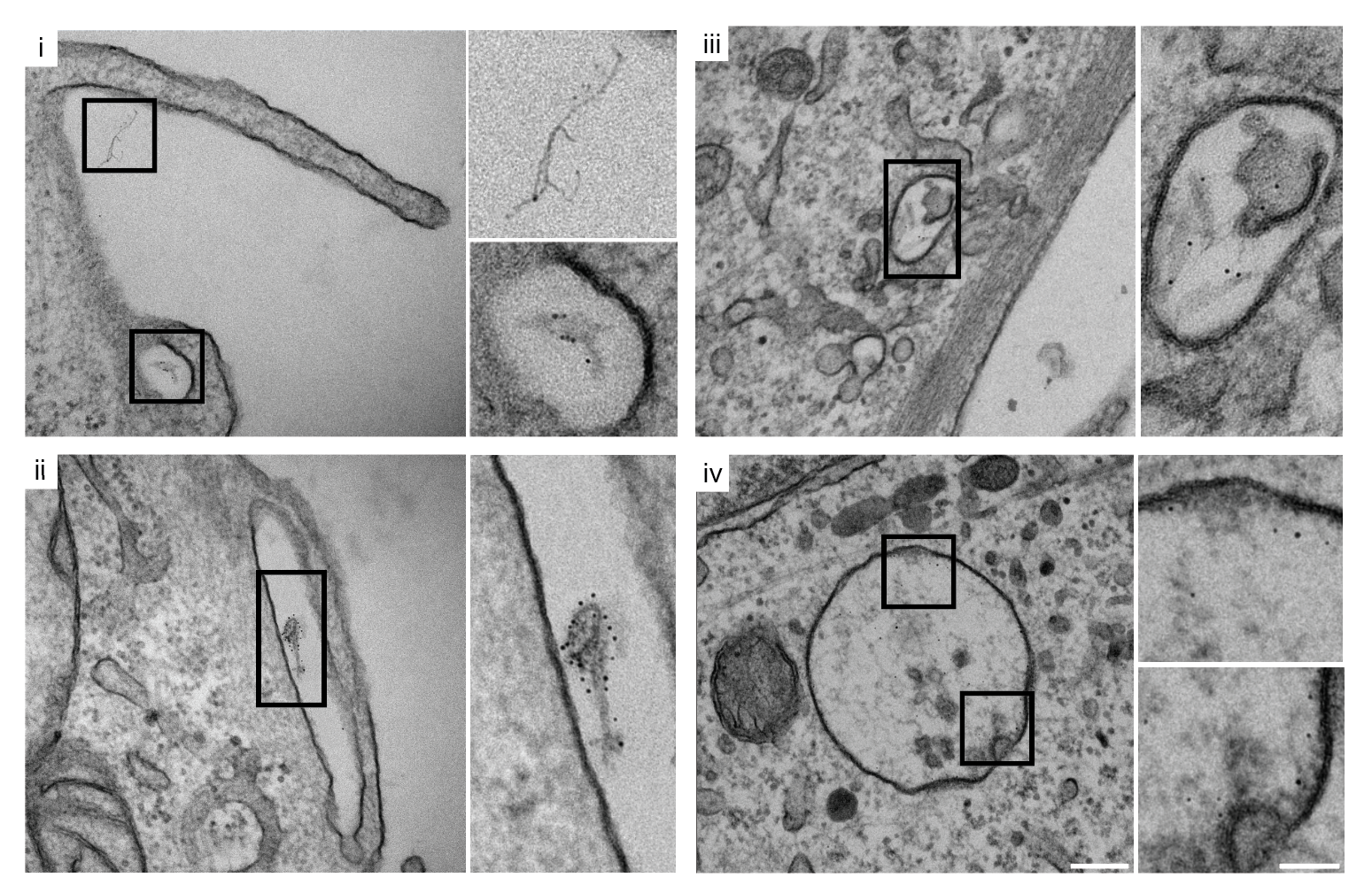
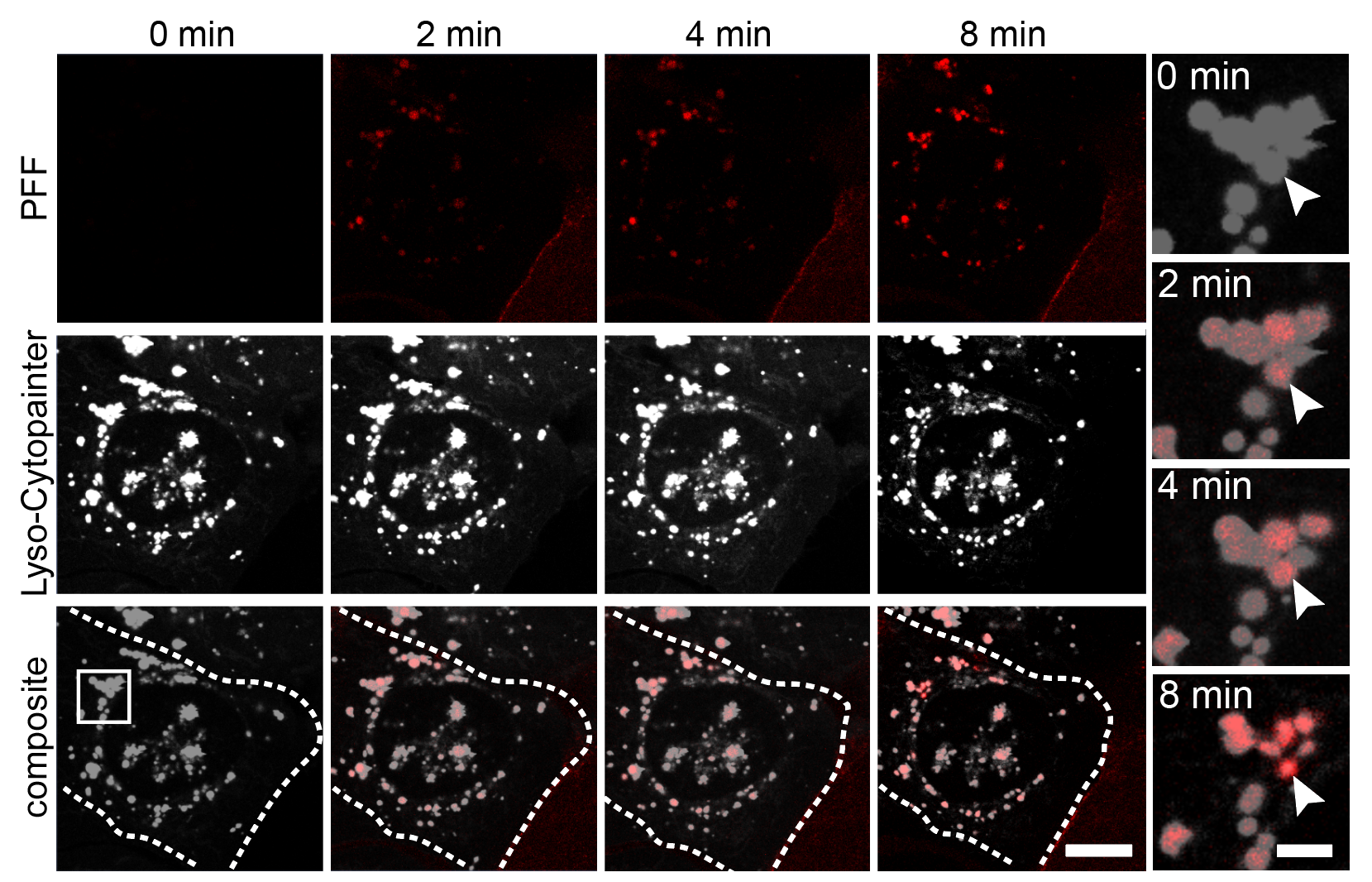
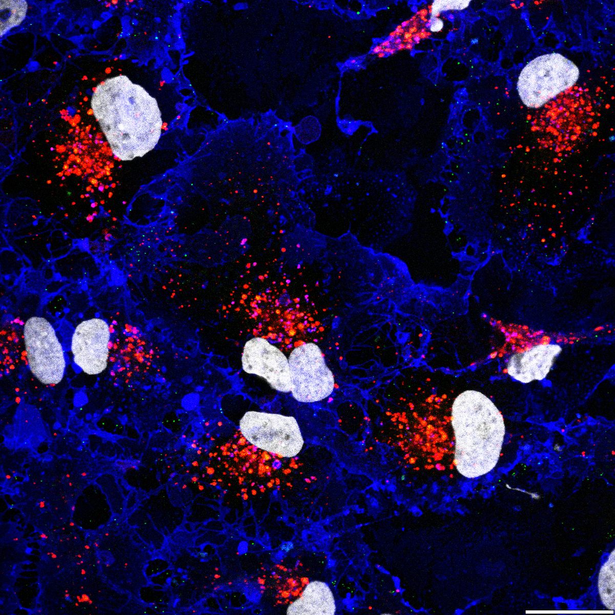
Using purified SARS-CoV-2 spike glycoprotein, lentiviral pseudotyping, and live-cell confocal microscopy, I demonstrated that SARS-CoV-2 relies on clathrin-mediated endocytosis (CME) for cell entry — establishing that viral RNA delivery to the cytosol occurs from within the endosomal lumen. This work is directly relevant to understanding how nucleic acid cargoes exploit endocytic machinery to access intracellular compartments, and how disrupting CME can modulate cargo delivery efficiency.

Experimental Approach

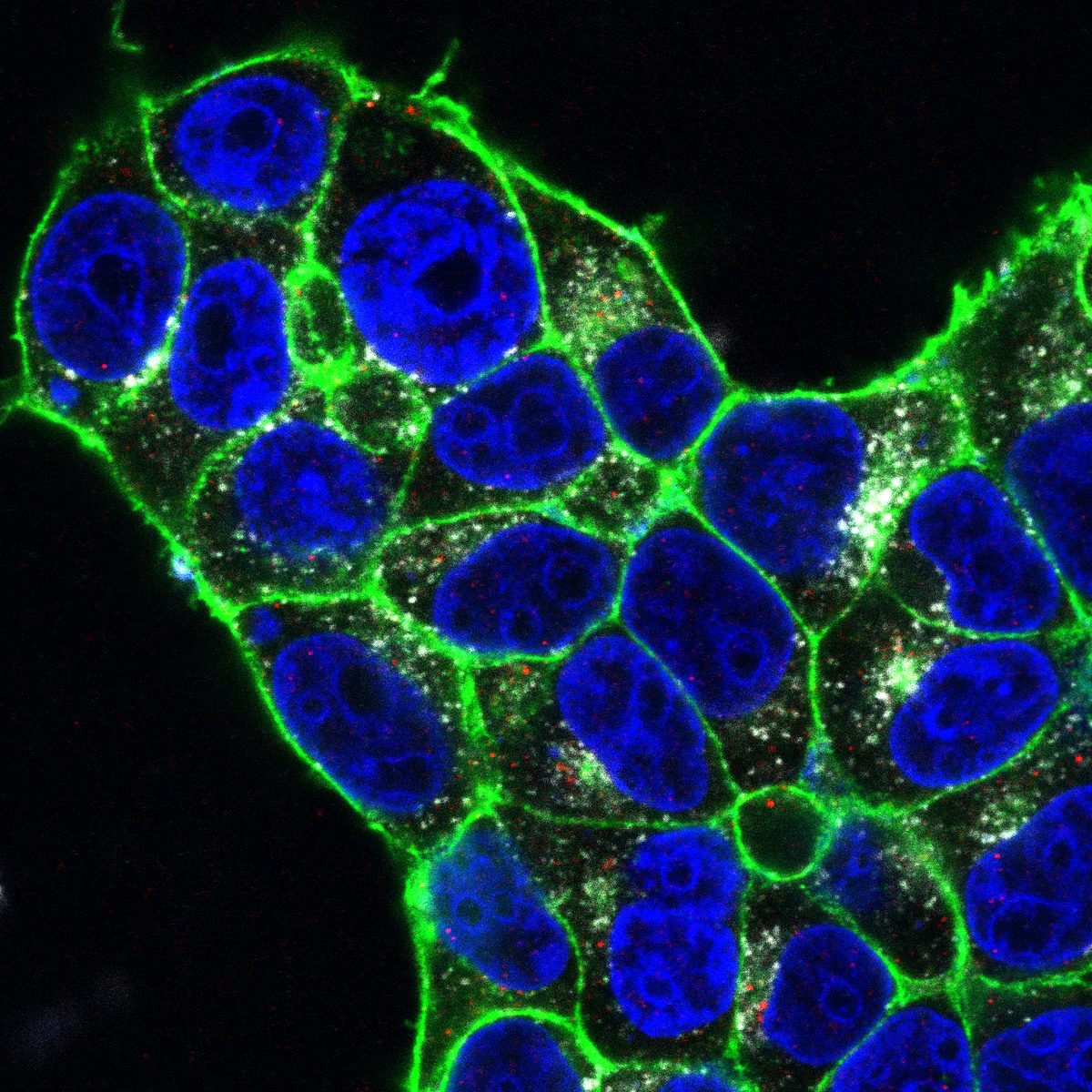
Cells expressing ACE2 (HEK-293T-ACE2, Vero, Calu-3) were incubated with purified His6-tagged spike protein or spike-pseudotyped lentivirus. Uptake was tracked by confocal microscopy at defined time points (2, 5, 15, 30 min). CME dependence was assessed via clathrin heavy chain (CHC) siRNA knockdown (~95% KD efficiency), pharmacological inhibition (Pitstop 2, Dynasore), and colocalization with clathrin/AP-2 markers.

🔬
Spike Protein Internalization via CME
CONFOCAL COLOCALIZATION siRNA KNOCKDOWN
Purified spike glycoprotein rapidly colocalized with clathrin and AP-2 at the plasma membrane within 2–5 minutes of exposure. CHC knockdown in Vero cells reduced spike internalization by 72%, and blocked pseudoviral infectivity by >80%. These findings confirmed that CME is the dominant entry route and that transfer of viral RNA to the cytosol requires transit through the endosomal system — a finding with direct implications for designing endosomal escape strategies for nucleic acid cargo.
Multi-Cell-Type Delivery Panel
HEK-293T-ACE2 VERO CALU-3 CACO-2
CME-dependent entry was validated across four cell lines representing diverse tissue origins (kidney, lung epithelial, intestinal epithelial), confirming that the pathway is not cell-type restricted. Each line was screened with pharmacological inhibitors and genetic knockdown to benchmark uptake efficiency — a workflow directly transferable to screening nucleic acid delivery vehicles across target cell panels.
Confocal: SARS-CoV-2 spike protein internalization via clathrin pathway
Confocal imaging of SARS-CoV-2 spike protein (green) internalization. Colocalization with clathrin confirms CME-dependent entry.
ACE2 receptor and spike protein colocalization on HEK-293T-ACE2 cells
ACE2 receptor engagement by spike protein on HEK-293T-ACE2 cells. Internalization reduced with PIKfyve and CSNK2 inhibitors.
Calu-3 epithelial cells with CK2 marker and spike puncta
Calu-3 airway epithelial cells showing spike puncta and CK2 marker. CSNK2 inhibition suppressed uptake.
Spike protein uptake at 15 minutes with DAPI nuclear stain
15-minute time point: spike puncta accumulation relative to DAPI nuclear counterstain across treatment conditions.

Quantitative CME Internalization Data

Data represent mean ± SEM from 3 independent experiments, n ≥ 200 cells per condition. Spike internalization quantified by intracellular fluorescence intensity normalized to surface-bound signal.

ConditionCell LineSpike Uptake (% Control)CHC Coloc. (Pearson's R)Pseudoviral Infectivity (%)
Vehicle controlHEK-293T-ACE2100 ± 6.20.78 ± 0.04100 ± 5.8
CHC siRNA (95% KD)HEK-293T-ACE228.4 ± 4.10.12 ± 0.0318.6 ± 3.2
Vehicle controlVero100 ± 7.40.74 ± 0.05100 ± 6.1
CHC siRNA (95% KD)Vero31.2 ± 5.30.14 ± 0.0422.1 ± 4.7
Pitstop 2 (30 μM)Vero42.6 ± 6.80.21 ± 0.0635.4 ± 5.9
Dynasore (80 μM)Vero38.1 ± 5.50.18 ± 0.0529.7 ± 4.3
Vehicle controlCalu-3100 ± 8.10.71 ± 0.06100 ± 7.2
Vehicle controlCaco-2100 ± 5.90.76 ± 0.04100 ± 4.8
−72%
Spike uptake with CHC knockdown
>80%
Pseudoviral infectivity blocked
4
Cell lines validated
R = 0.78
Spike–clathrin colocalization

Bayati, Kumar, Francis & McPherson. SARS-CoV-2 infects cells following viral entry via clathrin-mediated endocytosis. J. Biol. Chem. 2021; 296:100306.

02

Macropinocytic Cargo Uptake — Rapid, Clathrin-Independent Internalization

Not all cargo enters cells via CME. Using confocal, super-resolution (STED), and electron microscopy with immunogold labeling, I discovered that α-synuclein preformed fibrils (PFFs) bypass the classical endosomal pathway entirely and undergo rapid macropinocytic internalization, reaching lysosomes within 2 minutes of exposure. This work overturned the prevailing model of fibril entry and revealed a high-capacity, clathrin-independent uptake route — a pathway increasingly recognized as relevant for the delivery of large nucleic acid complexes and lipid nanoparticles.

Key Findings

2-Minute Lysosomal Delivery
STED LIVE-CELL CONFOCAL IMMUNOGOLD EM
Fluorescently labeled PFFs colocalized with LysoTracker and LAMP1 within 2 minutes of addition to SH-SY5Y and iPSC-derived dopaminergic neurons — far faster than any CME-dependent pathway could achieve. STED nanoscopy confirmed fibrils within the lysosomal lumen, not merely associated with lysosomal membranes. This ultrarapid delivery bypassed EEA1+ early endosomes and Rab11+ recycling endosomes entirely.
🔄
Pathway Dissection: CME vs. Macropinocytosis
siRNA PHARMACOLOGICAL INHIBITORS ACTIN/RAC1
Despite ~95% CHC knockdown efficiency, PFF uptake was completely unperturbed, ruling out CME. In contrast, macropinocytosis inhibitors EIPA, Latrunculin A/B, and Rac1 inhibition blocked uptake by 60–85%. PFFs induced actin- and Rac1-rich membrane ruffles — the hallmark precursors of macropinosomes. This pharmacological and genetic dissection framework is directly applicable to characterizing the entry routes of lipid nanoparticles, exosomes, and other nucleic acid delivery vehicles.
💎
Immunogold EM — Ultrastructural Cargo Tracking
NANOGOLD LABELING TEM SUBCELLULAR RESOLUTION
I developed a nanogold conjugation protocol for labeling α-synuclein fibrils, enabling antibody-free ultrastructural tracking by TEM. Gold-labeled fibrils were visualized at the plasma membrane during macropinosome formation, inside macropinosomes, and within lysosomes — providing direct spatial evidence of the entire internalization pathway at nanometer resolution. This technique is adaptable to tracking any conjugatable cargo, including labeled nucleic acids and nanoparticles.
EM panels: Nanogold-labeled fibrils at plasma membrane, macropinosomes, and lysosomes
TEM panels (i–iv): Nanogold-labeled fibrils at the plasma membrane during macropinocytosis initiation, within macropinosomes, and inside lysosomes.
Confocal: Rapid fibril uptake and lysosomal colocalization within 2 minutes
Confocal: PFF488 colocalization with LysoTracker within 2 min of exposure, confirming ultrarapid lysosomal delivery independent of early endosomes.
PFF-488 colocalized with LysoTracker and WGA membrane staining
PFF488 (green) colocalized with LysoTracker (red) and WGA membrane stain (blue). Demonstrates cargo reaching the lysosomal lumen while plasma membrane integrity is maintained.

Quantitative Pathway Dissection Data

PFF internalization quantified by intracellular fluorescence at 15 min. Mean ± SEM, n ≥ 150 cells per condition, 3 independent experiments. Inhibitor concentrations: EIPA 50 μM, LatA 0.5 μM, LatB 10 μM.

ConditionTargetPFF Uptake (% Vehicle)EEA1 Coloc. (R)LAMP1 Coloc. (R)Pathway
Vehicle control100 ± 5.40.08 ± 0.020.82 ± 0.03Macropinocytosis
CHC siRNA (95% KD)CME97.2 ± 6.10.07 ± 0.030.80 ± 0.04No effect
EIPA (50 μM)Na+/H+ exchanger22.4 ± 3.80.05 ± 0.020.31 ± 0.06Blocked
Latrunculin A (0.5 μM)Actin polymerization15.1 ± 2.90.04 ± 0.020.22 ± 0.05Blocked
Latrunculin B (10 μM)Actin polymerization18.6 ± 3.40.06 ± 0.030.28 ± 0.07Blocked
Rac1 inhibitorRac1 GTPase38.2 ± 5.70.07 ± 0.020.42 ± 0.05Blocked
Dynasore (80 μM)Dynamin88.4 ± 7.20.09 ± 0.030.76 ± 0.04Minimal effect
2 min
Time to lysosomal delivery
−85%
Uptake with LatA (actin block)
R = 0.82
PFF–LAMP1 colocalization
0%
CME contribution (CHC KD)

Bayati et al. Rapid macropinocytic transfer of α-synuclein to lysosomes. Cell Reports 2022; 40(3):111102.

03

Endosomal Trafficking, Subcellular Routing & Compartment Fate

A central question in cargo delivery is: once internalized, where does the cargo go? Using time-resolved colocalization with compartment-specific markers (EEA1, Rab5, Rab7, Rab11, LAMP1/2, CD63), I mapped the endosomal itinerary of multiple cargo classes. This work revealed that different cargoes follow distinct routing programs — a principle that governs whether internalized nucleic acids reach late endosomes (for escape) or are rapidly shunted to lysosomes (for degradation).

Compartment Mapping Across Cargo Classes

CargoEntry RouteEEA1+ (5 min)Rab7+ (15 min)LAMP1+ (30 min)Terminal Compartment
SARS-CoV-2 SpikeCME0.64 ± 0.050.72 ± 0.040.81 ± 0.03Late endosome → lysosome
α-Synuclein PFFsMacropinocytosis0.08 ± 0.020.34 ± 0.060.82 ± 0.03Direct to lysosome
EGF (CME control)CME0.81 ± 0.030.76 ± 0.040.68 ± 0.05Early → late endo → lysosome
Transferrin (recycling)CME0.84 ± 0.030.22 ± 0.050.09 ± 0.03Recycling endosome → PM
Dextran (10 kDa)Macropinocytosis0.12 ± 0.040.41 ± 0.060.78 ± 0.04Direct to lysosome
🔍
Endosomal Escape Window Identification
KINETIC COLOCALIZATION pH-SENSITIVE PROBES
By comparing the kinetic profiles of CME-dependent vs. macropinocytic cargoes, I identified a 5–15 minute window where CME-internalized cargo resides in mildly acidic late endosomes (pH ~5.5–6.0) before transfer to lysosomes. This window represents the critical period for endosomal escape of nucleic acid cargoes. In contrast, macropinocytic cargoes bypass this intermediate entirely, reaching lysosomes within 2 minutes — suggesting that delivery vehicles exploiting macropinocytosis may require alternative escape mechanisms or lysosomal disruption strategies.
💬
In Situ Subcellular Localization Across Cell Types
iPSC-DERIVED NEURONS SH-SY5Y HEK-293T U2OS PRIMARY GLIA
Subcellular routing was validated across 8+ cell types including iPSC-derived dopaminergic neurons, primary human astrocytes, and immortalized lines (SH-SY5Y, HEK-293T, U2OS, Hap1, Vero, CHO). Cell-type-dependent variations in trafficking kinetics were quantified, revealing that primary neurons route cargo to lysosomes 40% faster than immortalized lines — a key consideration for RNA delivery assay development.
5–15 min
Endosomal escape window (CME cargo)
8+
Cell types profiled for trafficking
6
Compartment markers used
04

Quantitative Internalization Kinetics & Dose-Response Profiling

For delivery assay development, quantitative kinetic parameters are essential. I developed plate-reader- and imaging-based workflows to generate dose-response curves, time-course profiles, and half-maximal uptake constants for diverse cargoes across cell types.

Time-Course Internalization Profiles

Fluorescence-based internalization quantified at 0, 2, 5, 10, 15, 30, 60 min post-exposure. Acid-wash step removes surface-bound cargo; remaining signal = intracellular. Mean ± SEM, n = 4.

CargoCell Typet1/2 Uptake (min)Max Uptake (%)Plateau (min)IC (nM or μg/mL)
Spike protein (5 μg/mL)HEK-293T-ACE24.2 ± 0.692 ± 4~20EC50 1.8 μg/mL
Spike protein (5 μg/mL)Vero5.1 ± 0.888 ± 5~25EC50 2.4 μg/mL
α-Syn PFFs (2 μg/mL)SH-SY5Y1.4 ± 0.396 ± 3~5EC50 0.8 μg/mL
α-Syn PFFs (2 μg/mL)iPSC-DA neurons1.1 ± 0.298 ± 2~3EC50 0.6 μg/mL
EGF (100 ng/mL)HEK-293T3.8 ± 0.594 ± 3~15EC50 42 nM
Transferrin (25 μg/mL)HEK-293T2.6 ± 0.491 ± 4~10EC50 8.4 μg/mL
Cargo Internalization Kinetics — Multi-Pathway Comparison
Intracellular fluorescence (acid-wash corrected) over time. Mean ± SEM, n = 4. PFFs show ultrarapid uptake vs CME-dependent cargoes.
PFF internalization plateaus by 5 min (macropinocytosis), while spike/EGF follow slower CME kinetics.

Dose-Response: Cargo Concentration vs. Uptake Efficiency

Cargo0.1×0.5×1× (Standard)Saturable?
Spike protein12 ± 354 ± 6100 ± 5142 ± 8168 ± 11Yes (receptor-limited)
α-Syn PFFs18 ± 462 ± 5100 ± 4186 ± 9324 ± 15No (fluid-phase)
EGF8 ± 248 ± 5100 ± 4128 ± 7139 ± 9Yes (receptor-limited)
Dextran 10 kDa14 ± 356 ± 6100 ± 5198 ± 10412 ± 18No (fluid-phase)
1.1 min
t1/2 uptake, PFFs in iPSC-DA neurons
EC50
Determined for all cargo/cell combinations
7
Time points per kinetic curve
05

Host Kinase Targets Controlling Endosomal Trafficking & Cargo Fate

Building on the mechanistic entry work, I contributed to two studies identifying druggable host kinases that control endosomal trafficking and cargo fate — PIKfyve and CSNK2. Both kinases regulate the endolysosomal system at nodes directly relevant to nucleic acid delivery: PIKfyve governs endosome-to-lysosome maturation, while CSNK2 modulates clathrin-mediated entry and endosomal processing. Pharmacological inhibition of either kinase disrupted viral cargo trafficking, demonstrating that host-directed strategies can redirect or block cargo routing through the endosomal system.

🔑
PIKfyve — Endosome-to-Lysosome Gatekeeper
CHEMICAL PROBE VIRAL ENTRY ENDOSOMAL MATURATION
PIKfyve synthesizes PI(3,5)P2, a phospholipid essential for endosomal maturation and lysosomal function. Using a novel, highly selective chemical probe (compound 17) with a matched negative control, we showed that PIKfyve inhibition blocks both viral replication and viral entry mediated by CME. PIKfyve inhibitors caused cargo to accumulate in enlarged, vacuolar endosomes rather than reaching lysosomes — effectively trapping cargo in the endosomal escape-permissive compartment. This finding suggests PIKfyve modulation could enhance endosomal escape of nucleic acid payloads.
CSNK2 — Clathrin Pathway & Trafficking Modulator
SILMITASERTIB BROAD-SPECTRUM ANTIVIRAL CME REGULATION
Casein kinase 2 (CSNK2) phosphorylates key regulators of clathrin-mediated endocytosis and vesicular trafficking. Using the clinical-grade inhibitor silmitasertib, we demonstrated that CSNK2 inhibition suppressed SARS-CoV-2 entry and replication across multiple β-coronavirus strains. Confocal imaging showed that silmitasertib reduced spike protein internalization by >60% in Calu-3 and Caco-2 cells. CSNK2 represents a host kinase that tunes the efficiency of clathrin-dependent cargo delivery.

Pharmacological Disruption of Endosomal Trafficking

InhibitorTargetViral Entry (% Ctrl)Viral Replication (% Ctrl)Cargo Trapping?
Compound 17 (1 μM)PIKfyve32 ± 58 ± 2Yes — enlarged endosomes
Compound 30 (neg. ctrl)None (inactive)96 ± 694 ± 5No
Apilimod (1 μM)PIKfyve28 ± 45 ± 2Yes — enlarged endosomes
Silmitasertib (5 μM)CSNK238 ± 612 ± 3Partial
SGC-CK2-1 (1 μM)CSNK242 ± 718 ± 4Partial
PIKfyve
Endosome maturation gatekeeper
CSNK2
CME and trafficking modulator
−92%
Viral replication (PIKfyve inhib.)
Cargo trapping
Endosomal escape window extended

Drewry, Potjewyd, … Bayati … et al. Identification and utilization of a chemical probe to interrogate the roles of PIKfyve in the lifecycle of β-coronaviruses. J. Med. Chem. 2022; 65(19):12860–12882.

Yang, Dickmander, Bayati et al. Host kinase CSNK2 is a target for inhibition of pathogenic SARS-like β-coronaviruses. ACS Chem. Biol. 2022; 17(7):1937–1950.

06

Subcellular Localization & Delivery Assay Toolkit

Across all of the above projects, I developed and refined a comprehensive toolkit for characterizing cargo localization, endosomal escape, and delivery efficiency. These methods are directly transferable to nucleic acid delivery screening and optimization workflows.

Techniques & Readouts

📸
Imaging-Based
Confocal colocalization (Pearson's R, Manders' coefficients) with compartment markers (EEA1, Rab5, Rab7, Rab11, LAMP1/2, CD63, Galectin-3). STED super-resolution for sub-lysosomal cargo localization. Immunogold EM for nanometer-scale trafficking. Live-cell time-lapse for real-time tracking. LysoSensor/LysoTracker pH profiling. High-content automated imaging (Opera Phenix) for plate-based screening.
📊
Quantitative Readouts
Plate-reader fluorescence/luminescence for high-throughput internalization. Flow cytometry for single-cell uptake quantification. RT-qPCR for mRNA delivery and expression. Western blot for protein expression. ELISA / Luminex for cytokine profiling after cargo exposure. Acid-wash assays discriminating surface-bound from intracellular cargo.

Cell Type Experience Panel

All endocytosis and trafficking assays validated across a broad panel of mammalian cell types:

CategoryCell TypesSource
iPSC-derivedDopaminergic neurons, cortical neurons, astrocytes, microglia-like, cardiomyocytesPatient-derived & healthy control
Primary humanAstrocytes, PBMCs, T cells, monocytesDonor tissue
EpithelialCalu-3, Caco-2, Vero, HeLaLung, gut, kidney
Neuronal/neuroblastomaSH-SY5Y, U87, U251, Hap1Immortalized
Fibroblast/kidneyHEK-293T, HEK-293T-ACE2, CHOEngineered & parental
OtherU2OS, HMC3 (microglia), THP-1Immortalized
20+
Cell types used for endocytosis assays
3
Microscopy modalities (confocal, STED, EM)
6
Quantitative readout platforms

Cargo Delivery Assay Development Workflow

01
Cargo & Cell Selection
Label cargo (fluorescent, nanogold, or reporter). Select cell panel spanning primary, iPSC-derived, and immortalized lines. Establish dose and time-course parameters.
02
Entry Pathway Mapping
Pharmacological and genetic pathway dissection (CME, macropinocytosis, caveolae). siRNA/shRNA knockdown. Inhibitor panels (EIPA, Pitstop, Dynasore, LatA/B).
03
Trafficking & Localization
Time-resolved colocalization with compartment markers. Identify endosomal escape window. pH-sensitive probes. STED/EM for ultrastructural confirmation.
04
Functional Readout
RT-qPCR for mRNA expression. Flow cytometry for single-cell delivery. ELISA/Luminex for immune response. Plate-reader for high-throughput screening.