ESC
Movement Disorder Research

Parkinson's Disease & Dystonia

A consolidated view of my research on Parkinson's disease and dystonia: iPSC-derived dopaminergic, cortical, and striatal-adjacent neuron models, α-synuclein pathology, lysosomal dysfunction, CRISPR-edited disease lines (LRRK2, GBA, Parkin, SNCA), neuroinflammation, electrophysiology, quantitative biomarkers, and small-molecule / biologic neuroprotection — integrating mechanism and translation for movement disorders.

01

Movement Disorders — An Integrated Research Program

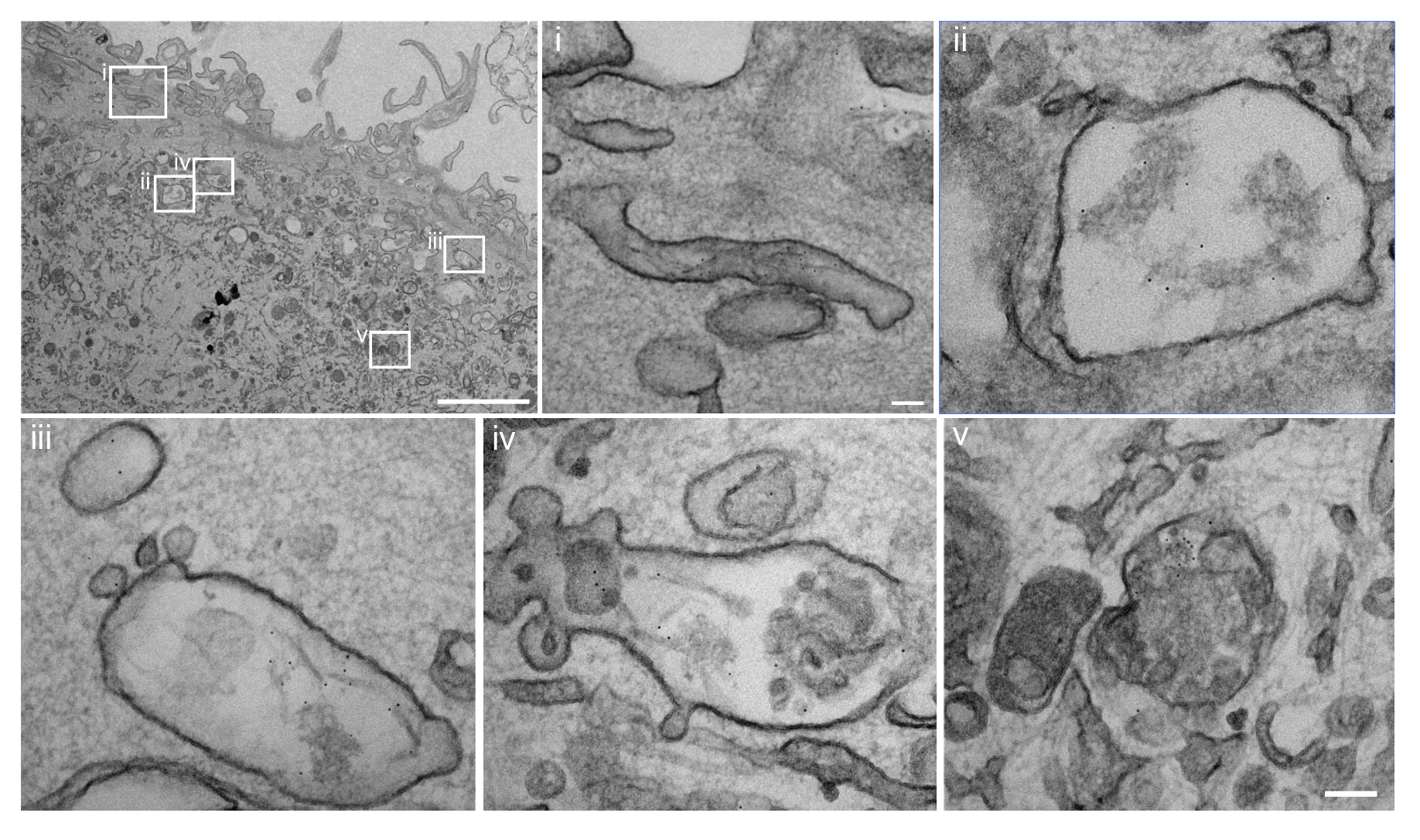
My work centers on the molecular and cellular biology of Parkinson's disease — α-synuclein pathology, lysosomal dysfunction, dopaminergic neuron vulnerability, and neuroinflammation — built on iPSC-derived human neuron platforms, CRISPR-edited disease lines, and quantitative multi-modal readouts. The same platform translates directly to dystonia and other basal-ganglia / motor-circuit disorders: iPSC differentiation into dopaminergic and cortical/striatal-projecting neurons, microelectrode-array (MEA) electrophysiology, calcium imaging, high-content confocal and electron microscopy, CRISPR knockout modeling, and mass-spectrometry proteomics. This page consolidates the core figures, assays, and findings from across the site that are directly relevant to evaluating my capabilities in Parkinson's disease and dystonia.

9
Patient / Donor iPSC Lines
20+
iPSC Lines Generated & Banked
4
PD-Relevant CRISPR KOs
4.2×
α-Syn Inclusion Increase (Dual-Hit)

Research Pillars

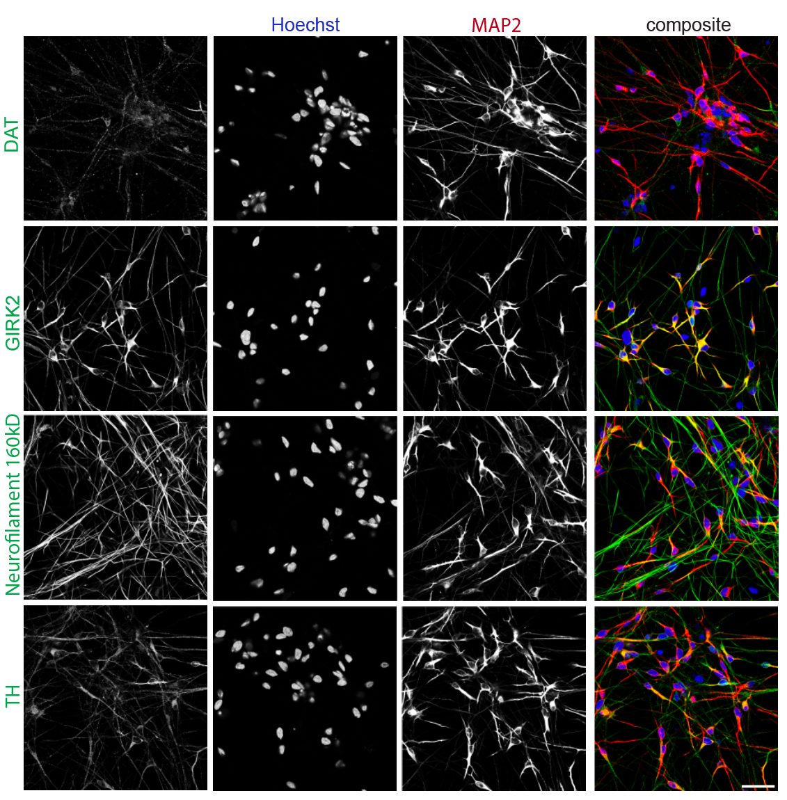
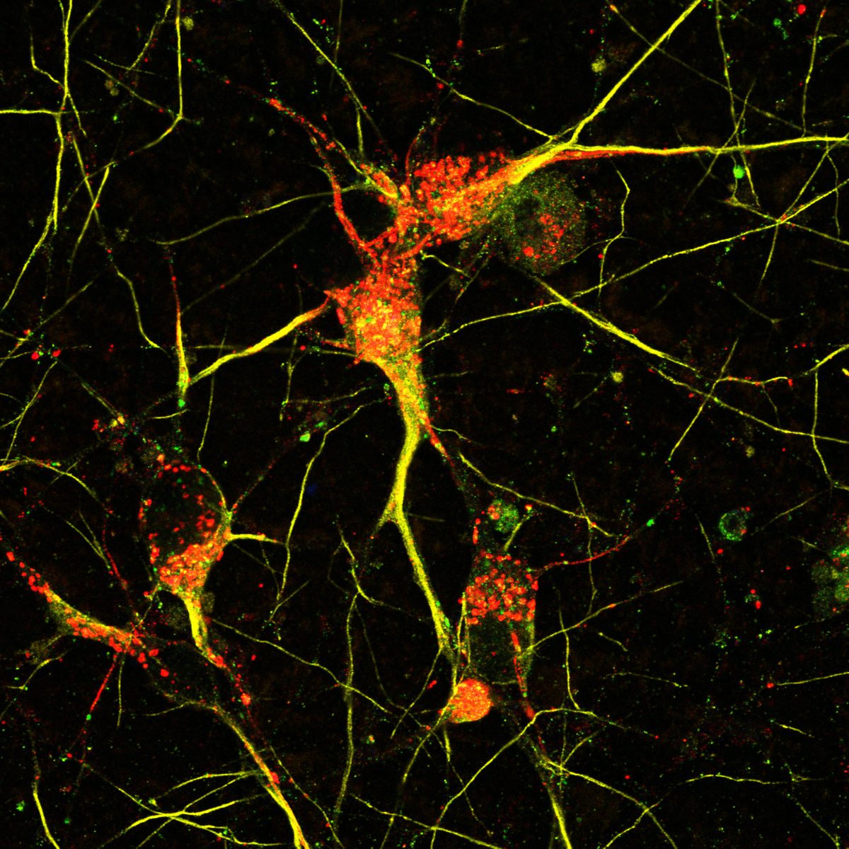
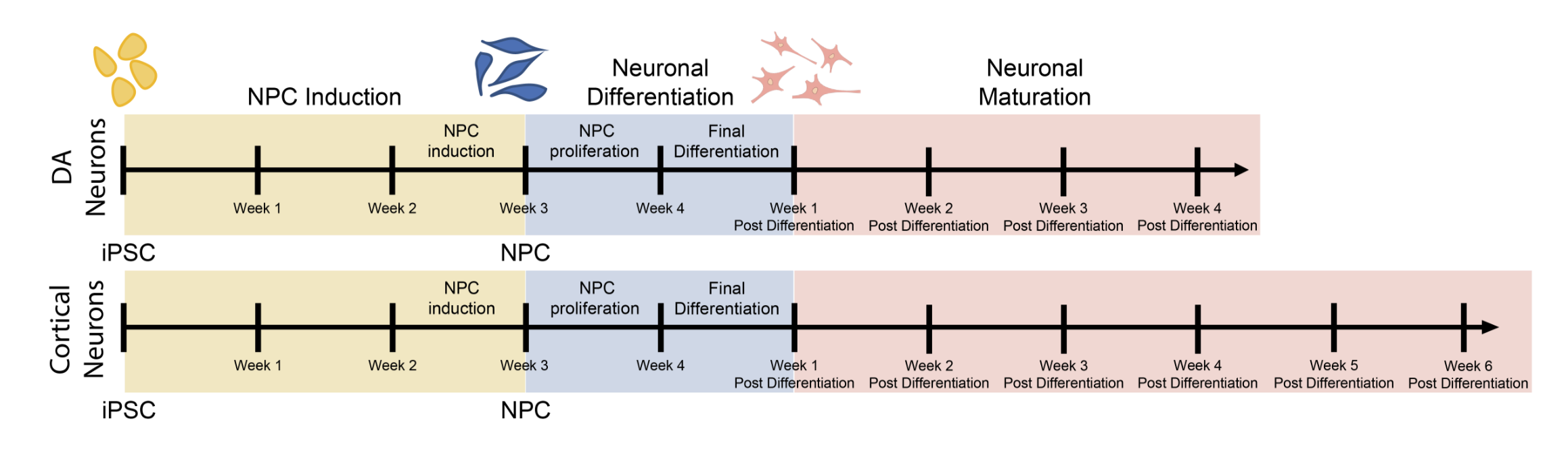
  • iPSC-derived midbrain dopaminergic & cortical neurons
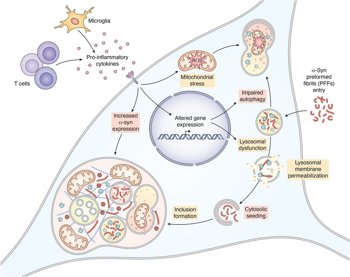
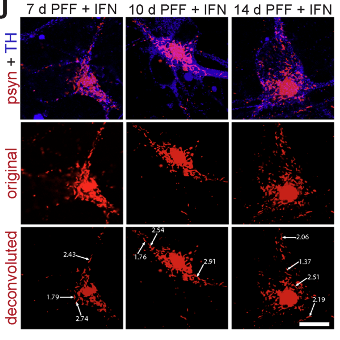
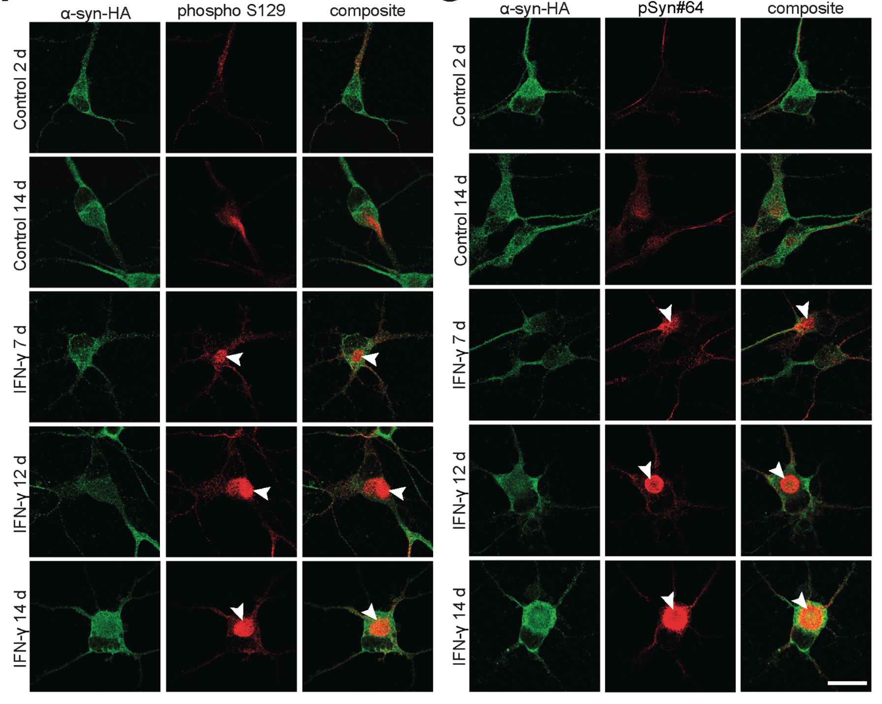
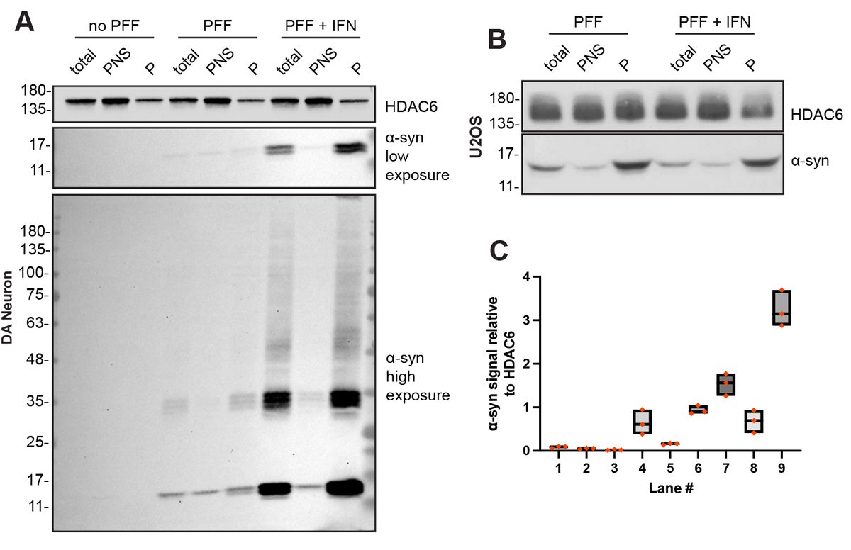
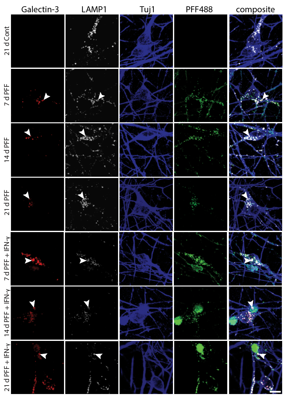
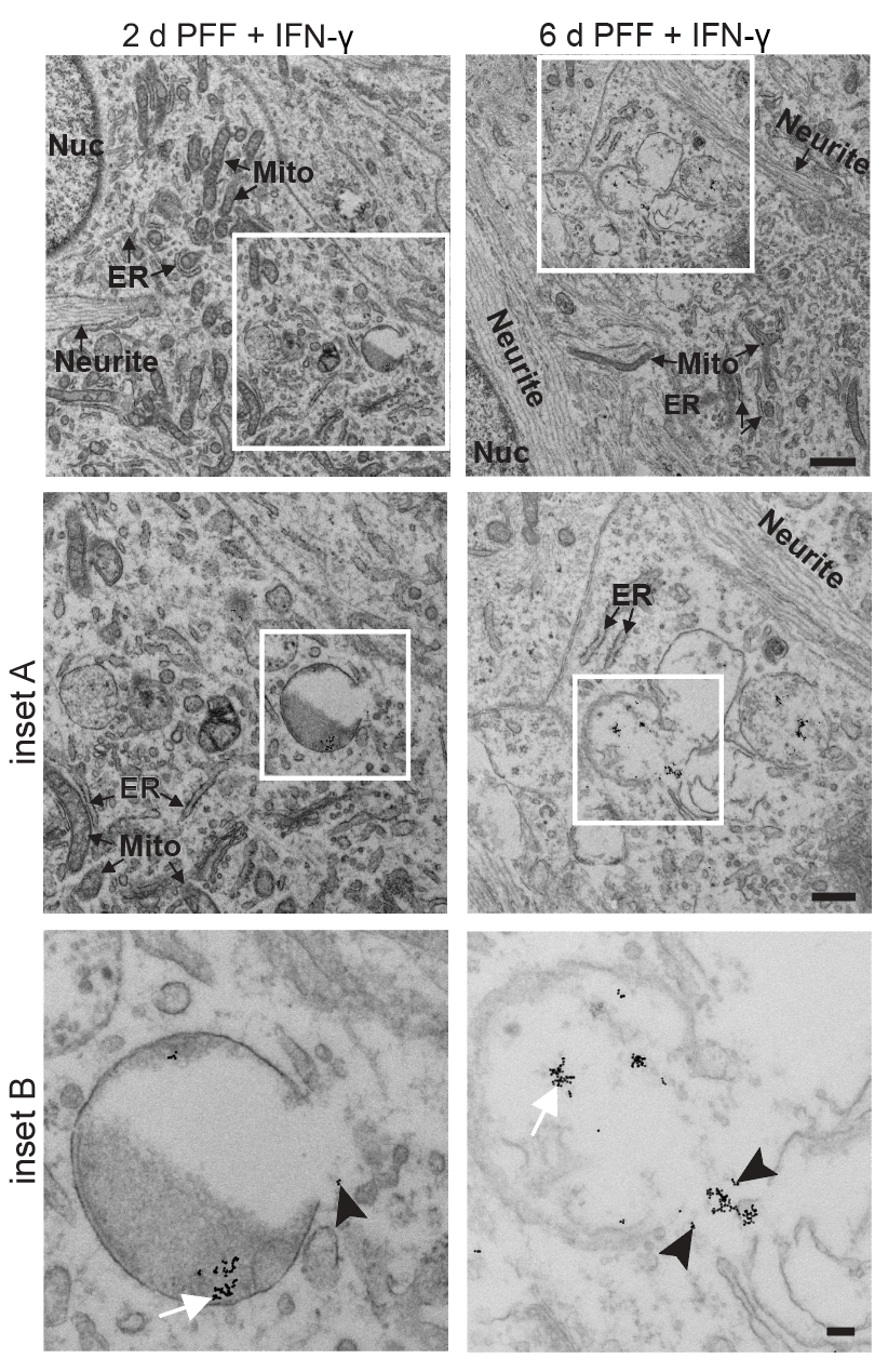
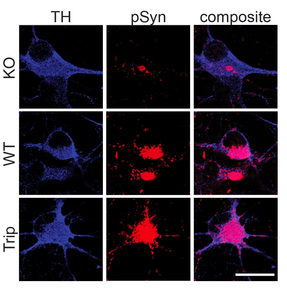
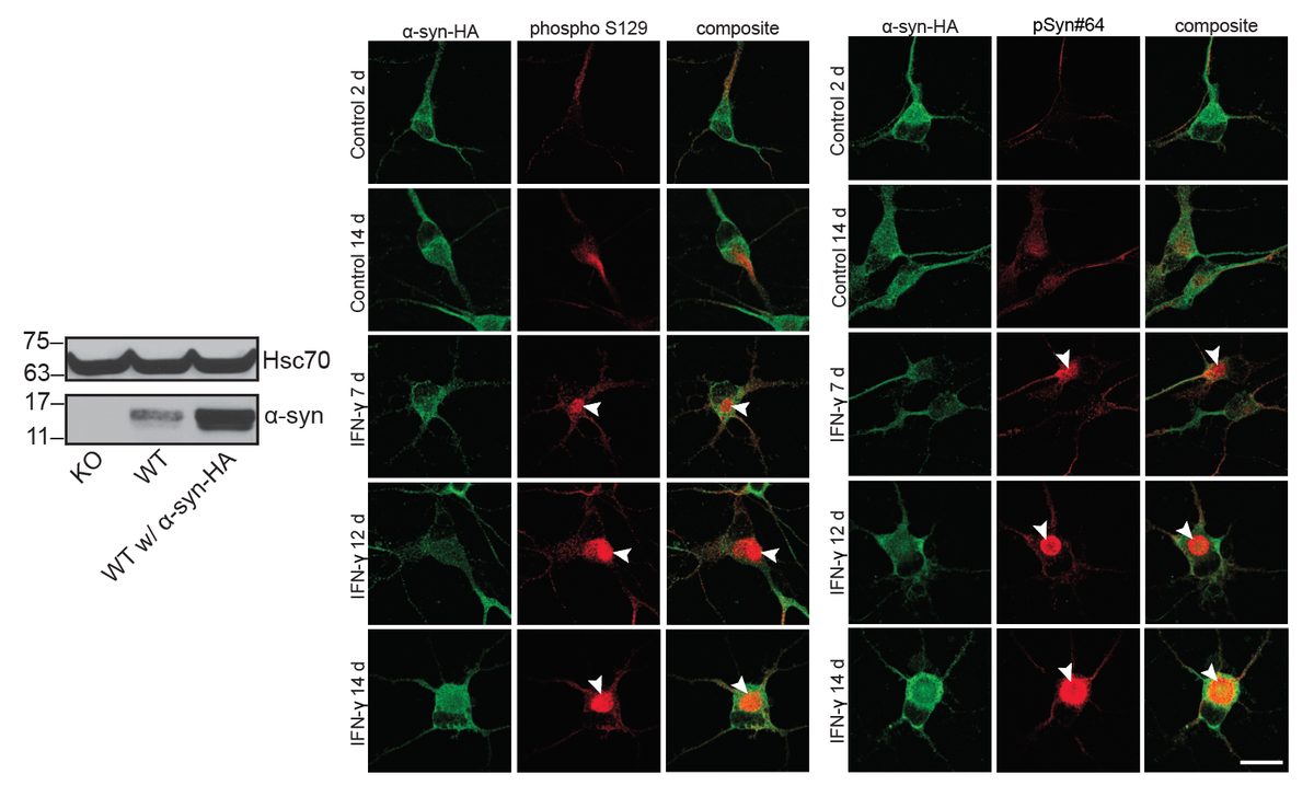
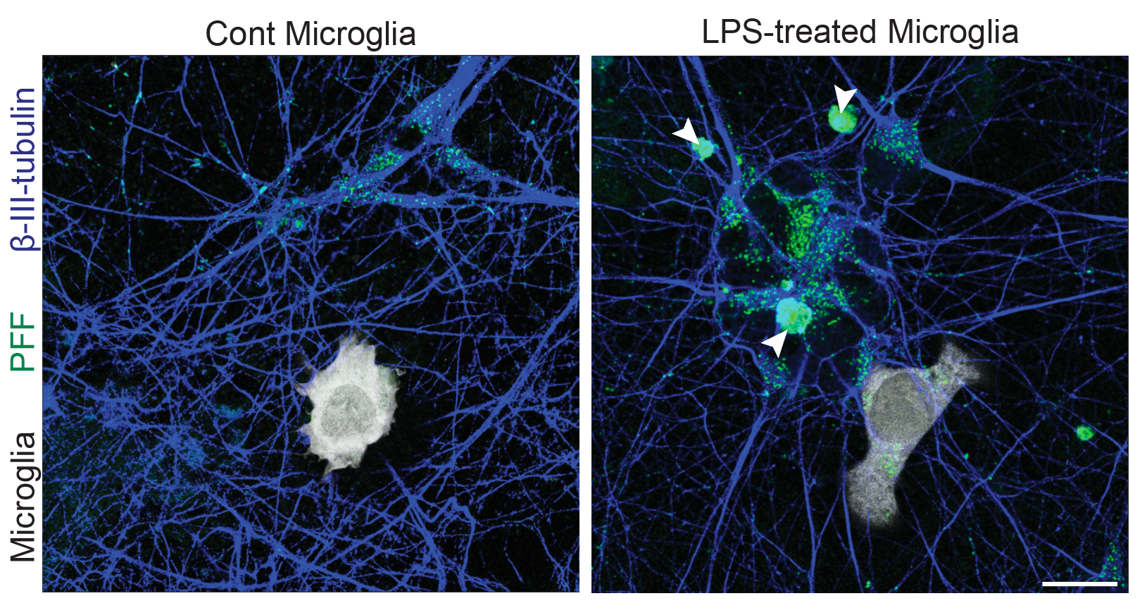
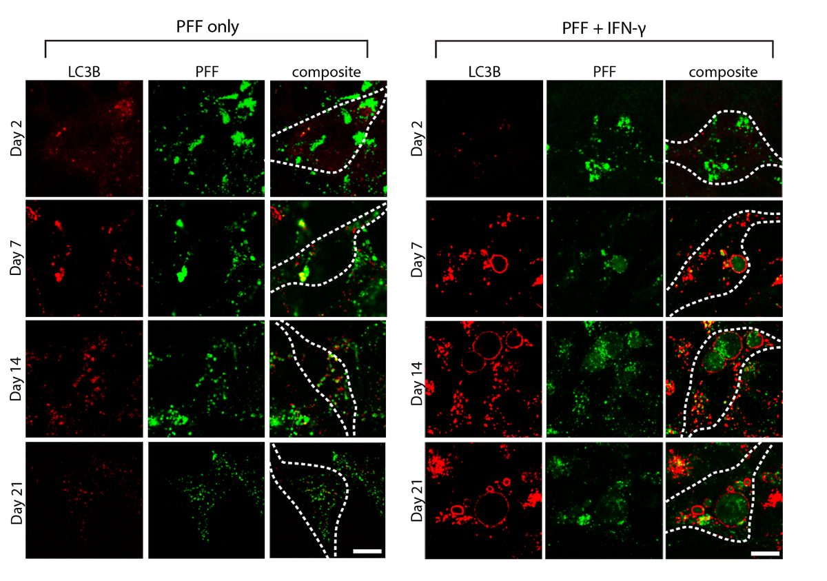
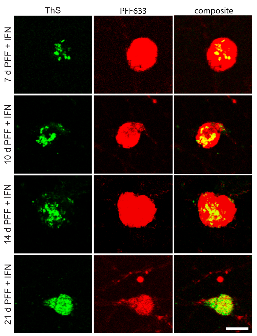
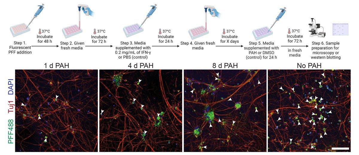
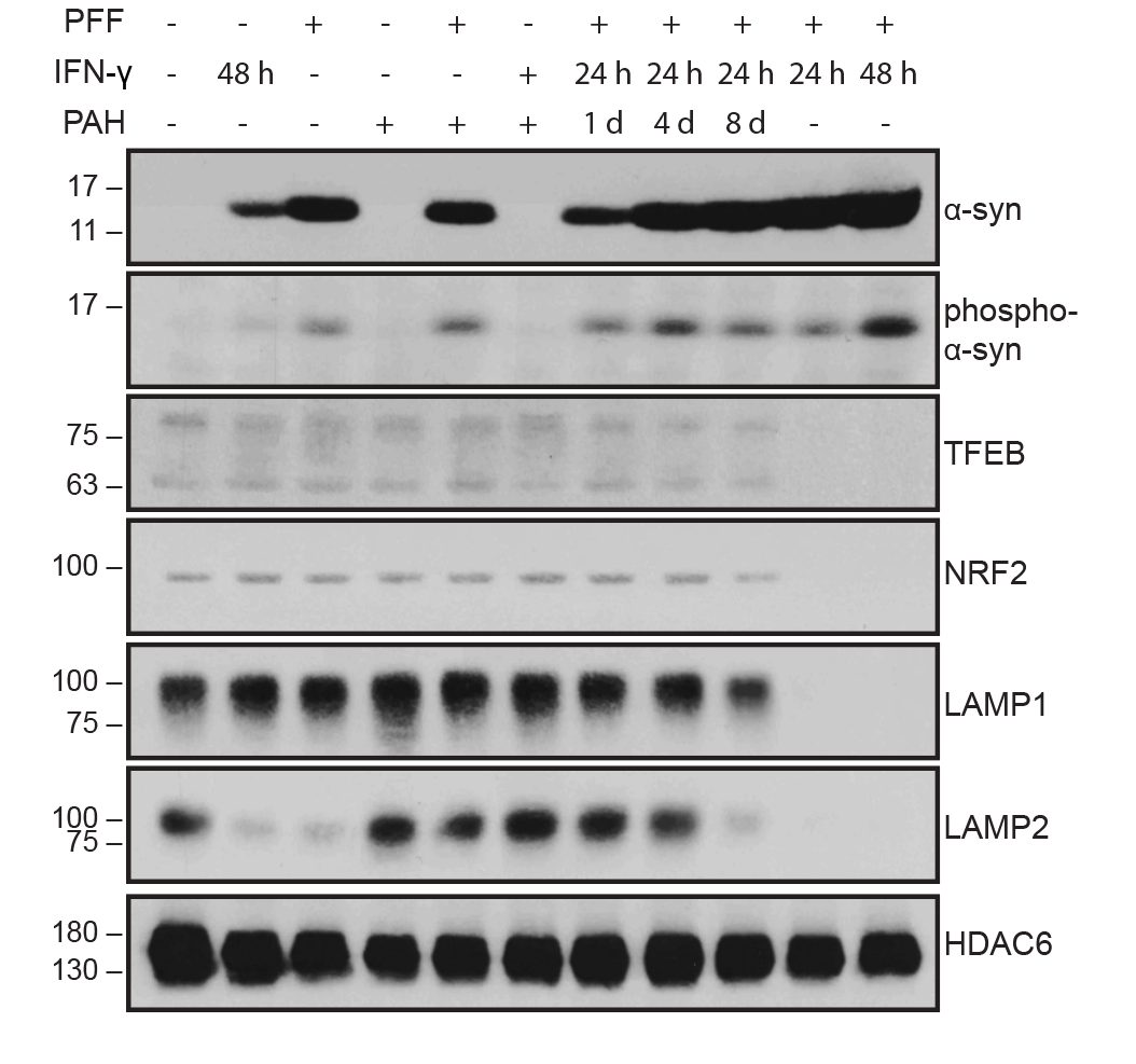
  • α-Synuclein PFF seeding & dual-hit Lewy-body modeling
  • CRISPR-edited disease lines (LRRK2, GBA, Parkin, SNCA)
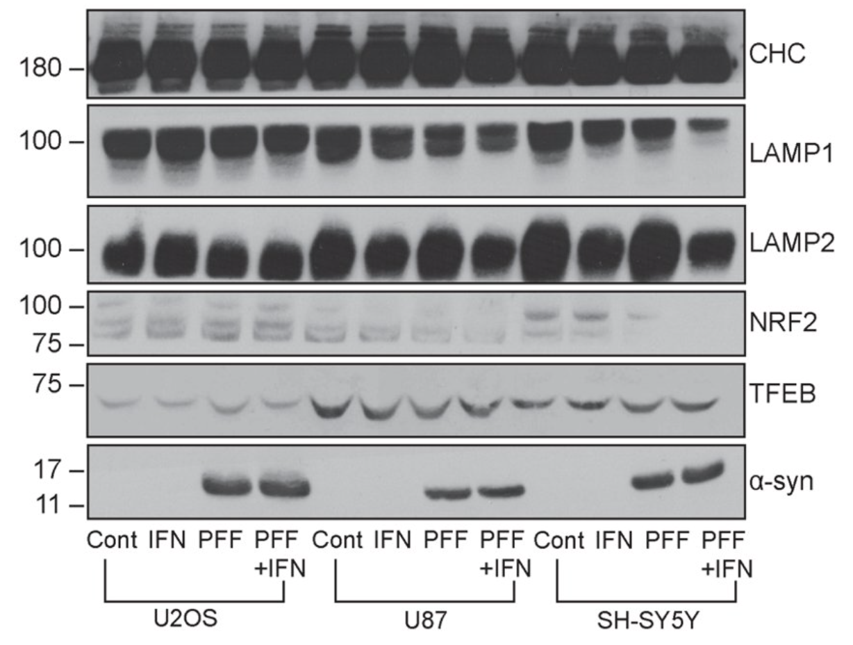
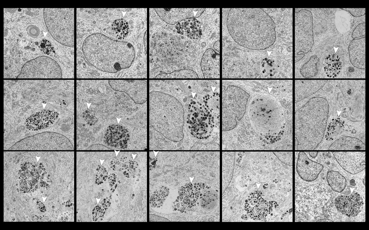
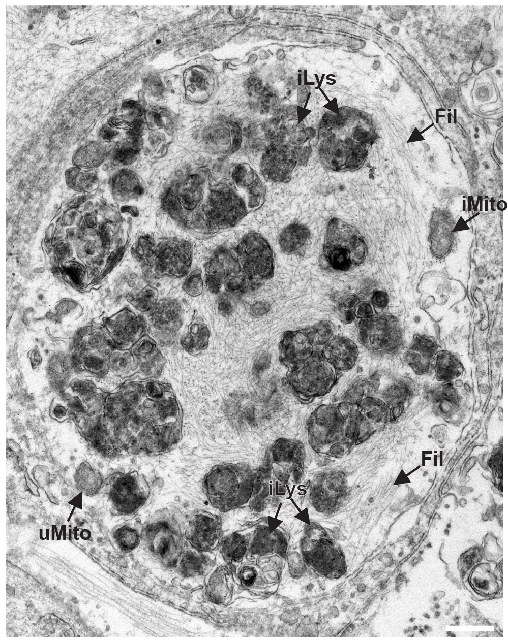
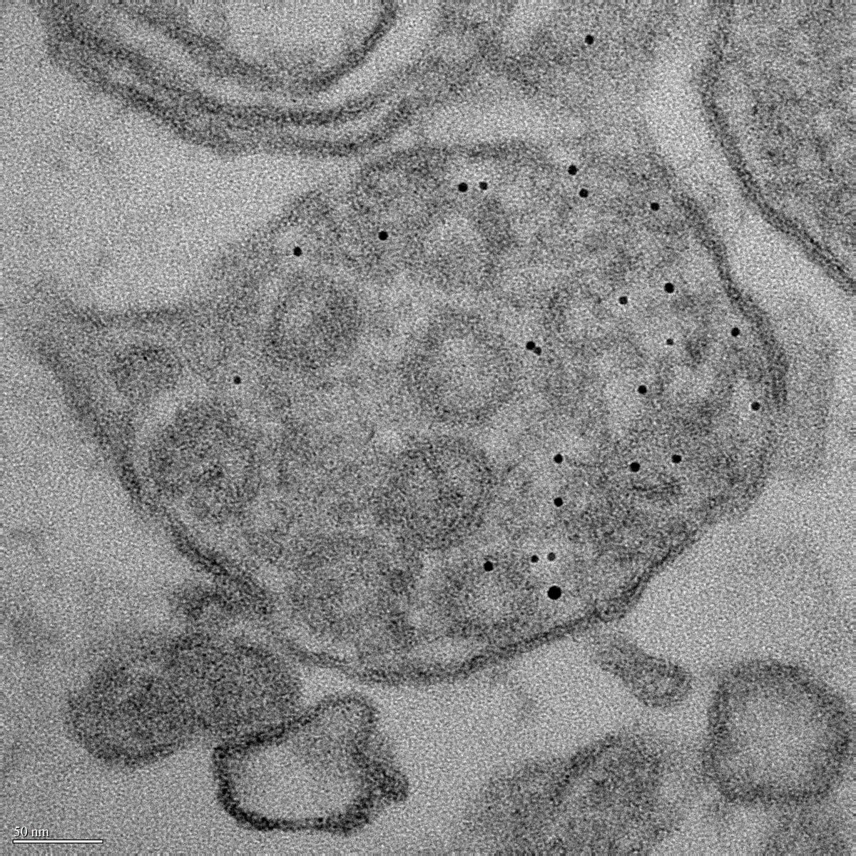
  • Lysosomal dysfunction & membrane permeabilization
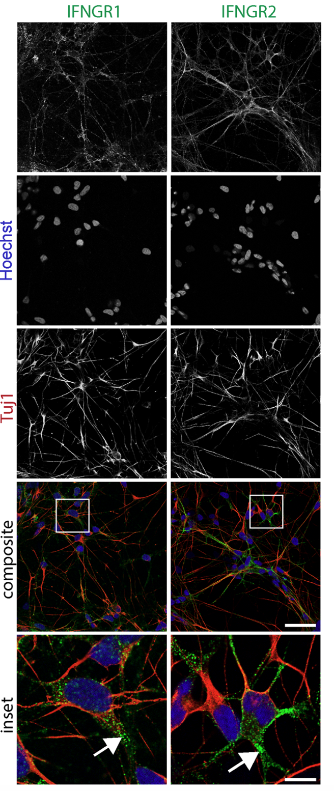
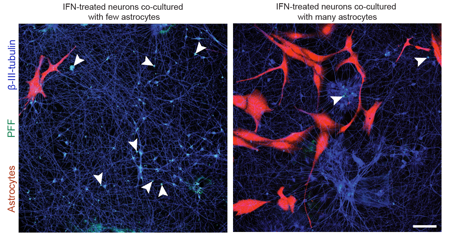
  • Neuroinflammation (IFN-γ, TNF-α, IL-1β) and microglial co-culture
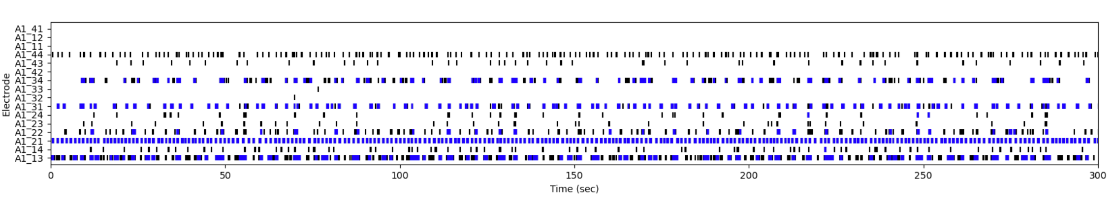
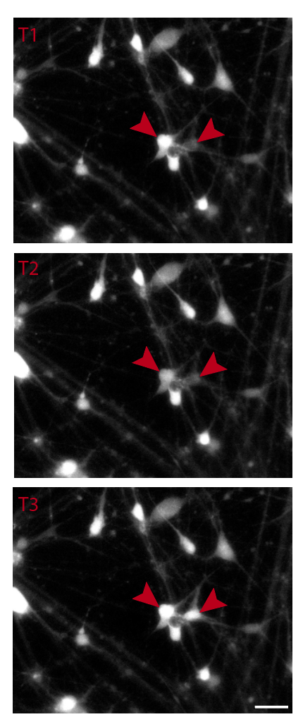
  • MEA electrophysiology & calcium imaging — circuit-level readouts for movement disorders
  • Parkin-agonist & lysosomal-rescue small-molecule discovery
  • Biomarker development: pS129, LAMP1/2, TFEB, NfL, HDAC6
02

Basal Ganglia Circuitry — PD vs Dystonia (Interactive)

The basal ganglia organize voluntary movement through the balance of a direct (movement-promoting) and an indirect (movement-inhibiting) pathway, modulated by dopamine from the substantia nigra pars compacta (SNc) and a fast-stop hyperdirect projection from motor cortex to the subthalamic nucleus (STN). Parkinson’s disease and dystonia are both classical basal-ganglia disorders yet produce opposite motor phenotypes — hypokinetic bradykinesia/rigidity/tremor versus hyperkinetic, sustained agonist–antagonist co-contraction. Click a state below to visualize pathway-specific dysfunction.

Basal ganglia circuit schematic Direct, indirect and hyperdirect pathways showing dysfunction in Parkinson's disease and dystonia. Motor Cortex M1 · SMA Striatum D1 GABA · SP / Dyn Striatum D2 GABA · Enk GPe Globus pallidus externa STN Subthalamic · Glu GPi / SNr Output nucleus · GABA Thalamus (VA / VL) Motor relay · Glu SNc Dopamine D1 (+) · D2 (−) !
Direct pathway (movement-promoting) Indirect pathway (movement-inhibiting) Hyperdirect (cortex → STN) Dopamine (SNc) excitatory inhibitory
Healthy Balance

Phasic SNc dopamine excites D1 (movement-promoting direct pathway) and inhibits D2 (movement-suppressing indirect pathway). Net GPi/SNr output gates thalamo-cortical excitation, permitting accurately scaled, appropriately selected voluntary movement. The hyperdirect cortico-STN projection supplies a fast stop signal for action cancellation.

  • Balanced direct ↔ indirect tone
  • Phasic dopamine shapes action selection
  • Surround inhibition at motor cortex preserves selectivity
Parkinson’s Disease — Hypokinetic

α-Synuclein Lewy pathology drives progressive loss of SNc dopaminergic neurons. Reduced D1 drive weakens the direct pathway; disinhibited D2-striatal activity over-suppresses GPe, which over-excites STN → GPi/SNr. The resulting excess GPi inhibition over-clamps thalamus and starves motor cortex of activating drive — manifesting as bradykinesia, rigidity, and resting tremor.

  • ↓ SNc dopamine (α-syn pathology)
  • ↑ STN firing → ↑ GPi output
  • ↓ Thalamo-cortical drive
  • Therapeutics: L-DOPA, DBS-STN/GPi, GBA / PARK2 / LRRK2 modulation
Dystonia — Hyperkinetic / Co-contraction

Heterogeneous monogenic etiologies (TOR1A/DYT1, GNAL, THAP1, KMT2B, SGCE, ATP1A3) converge on reduced GPi/SNr inhibitory output, producing thalamic disinhibition and aberrant, excessive cortical activation with loss of surround inhibition. Maladaptive plasticity at cortico-striatal synapses (abnormal LTP/LTD) reinforces the pattern, producing involuntary, sustained agonist–antagonist co-contraction and twisting postures.

  • ↓ GPi/SNr output → thalamic disinhibition
  • Loss of surround inhibition at cortex
  • Aberrant cortico-striatal plasticity (mal-LTP/LTD)
  • Therapeutics: DBS-GPi, anti-cholinergics, botulinum toxin, gene-specific (KMT2B, GNAL)
Fig. Interactive schematic of the basal-ganglia direct / indirect / hyperdirect loops in health, Parkinson’s disease, and dystonia. Click a state to recolor altered pathways and reveal node-level dysfunction badges.
03

Human iPSC Dopaminergic & Cortical Neuron Models

I have generated and banked over 20 iPSC lines, including nine patient/donor lines used for modeling Parkinson's disease pathology. Differentiation spans midbrain dopaminergic neurons (for PD modeling), cortical / excitatory neurons (for circuit studies relevant to both PD cognitive features and dystonia motor-cortex biology), astrocytes, and microglia-like cells for co-culture. Cell identity is confirmed by immunocytochemistry (TH, DAT, GIRK2, Neurofilament, TUJ1, MAP2), HPLC quantification of dopamine and metabolites, and MEA electrophysiology for spontaneous firing patterns and waveform analysis.

Identity & Quality Control

  • TH+ midbrain dopaminergic neurons (DAT, GIRK2, Neurofilament co-stain)
  • Cortical excitatory neurons (TUJ1, MAP2, VGLUT1)
  • HPLC dopamine & DOPAC quantification
  • MEA spontaneous activity & spike waveform analysis
  • Calcium imaging (Fluo-4) of spontaneous transients
  • Karyotyping, pluripotency panel, colony QC
iPSC-derived dopaminergic neuron ICC panel: DAT, GIRK2, Neurofilament, and TH co-staining confirming midbrain DA identity
Fig. iPSC-derived DA neuron identity — DAT, GIRK2, Neurofilament, and TH co-expression

Relevance to Dystonia

The same iPSC differentiation, MEA, and calcium-imaging pipelines are directly applicable to dystonia modeling — circuit-level firing-pattern abnormalities, paroxysmal burst activity, and co-contraction-like synchrony phenotypes are quantifiable on our MEA platform, and patient-derived lines carrying movement-disorder alleles (e.g., TOR1A/DYT1, GNAL, THAP1, KMT2B) can be differentiated into cortical and striatal-adjacent neurons for comparative electrophysiology and morphology. Where direct PD data is shown below, the underlying methodology — from differentiation to high-content imaging to CRISPR perturbation — transfers to dystonia models with minimal adaptation.

04

α-Synuclein Biology, PFF Seeding, & Lewy-like Inclusions

Sonicated recombinant α-synuclein preformed fibrils (PFFs) seed endogenous α-synuclein aggregation in iPSC-DA neurons. Fibrils are internalized primarily by RAC1-dependent macropinocytosis, bypass early endosomes, and reach lysosomes within two minutes of addition. Seeds recruit endogenous α-synuclein and template the formation of pS129-positive, Thioflavin-S-positive Lewy-like inclusions over 14–21 days. A dual-hit paradigm (PFF + IFN-γ) dramatically accelerates inclusion formation and models the inflammatory context of sporadic PD.

Schematic: dual-hit Parkinson's model combining α-synuclein PFFs and IFN-γ inflammation driving lysosomal dysfunction and Lewy body formation
Fig. Dual-hit Parkinson's model — PFFs + cytokine challenge → lysosomal dysfunction → Lewy-like pathology

Pharmacodynamic Biomarker Panel

Multi-target panel quantifying α-synuclein (pS129 & total), lysosomal proteins (LAMP1/LAMP2), autophagy flux (LC3B, p62), transcriptional regulation (TFEB), and aggresome machinery (HDAC6) across vehicle, dual-hit, and rescue conditions.

Pharmacodynamic Biomarker Panel
Dual-Hit PD Model (PFF + IFN-γ) with Drug Rescue
Inclusion Growth Over Time

Time-resolved phospho-synuclein (pSyn) inclusion growth in TH+ dopaminergic neurons exposed to PFF + IFN-γ over 7, 10, and 14 days. Deconvoluted panels with size measurements (µm) show progressive inclusion maturation and coalescence, providing quantitative morphometric data to benchmark therapeutic rescue.

pSyn + TH time course at 7d, 10d, 14d under PFF+IFN showing original and deconvoluted inclusions with size measurements
Fig. pS129 α-synuclein inclusion growth — deconvoluted morphometry at 7, 10, and 14 days post dual-hit
Orthogonal Antibody Validation

Confocal time course of α-synuclein-HA expressing neurons probed with two independent phospho-synuclein antibodies (phospho S129 and pSyn#64). Control neurons show diffuse cytoplasmic α-syn-HA signal; IFN-γ–treated neurons develop perinuclear pS129-positive inclusions by day 7 that mature through day 14 — validating the biomarker with orthogonal antibody approaches.

α-syn-HA with phospho S129 and pSyn#64 antibodies: control vs IFN-γ time course showing progressive inclusion formation from 2d to 14d
Fig. Phospho-synuclein biomarker validation — dual antibody time course (pS129 & pSyn#64)
Biochemical Isolation of Inclusions

Sequential extraction and subcellular fractionation reveal enrichment of α-synuclein and HDAC6 (aggresome marker) in the insoluble pellet under dual-hit conditions, biochemically confirming the confocal pathology.

Subcellular fractionation Western blots (soluble, membrane, pellet) showing α-synuclein and HDAC6 enriched in insoluble pellet
Fig. Sequential extraction — α-synuclein & HDAC6 enrichment in the insoluble pellet under dual-hit
05

Lysosomal Dysfunction, GBA & Fibril Trafficking

Lysosomal biology sits at the center of Parkinson's disease pathogenesis — and is increasingly implicated in movement-disorder biology more broadly. I developed a custom nanogold-PFF conjugation protocol, published in STAR Protocols (first author), that permits direct electron-microscopic visualization of fibril trafficking from the cell surface into lysosomes. Live-cell confocal (LysoTracker / LysoSensor) quantifies lysosomal number and pH; galectin-3 puncta report membrane permeabilization; and GBA-knockout iPSC neurons reveal heightened vulnerability to fibril-induced inclusions.

Western Blot Pharmacodynamic Panel

Multi-target Western blot across U2OS, U87, and SH-SY5Y cell lines under control, IFN-γ, PFF, and dual-hit conditions quantifies CHC (clathrin heavy chain), LAMP1, LAMP2, NRF2, TFEB, and α-synuclein in a single readout — capturing endocytic machinery, lysosomal integrity, oxidative stress, and aggregation state.

Western blot panel showing CHC, LAMP1, LAMP2, NRF2, TFEB, α-syn across U2OS, U87, SH-SY5Y cell lines
Fig. Pharmacodynamic Western — CHC, LAMP1, LAMP2, NRF2, TFEB, α-synuclein across cell lines
06

CRISPR-Edited iPSC Lines for PD & Movement-Disorder Biology

I have generated CRISPR-edited iPSC lines targeting major Parkinson's disease genes to dissect protein function and model familial forms of disease. The same workflow — ribonucleoprotein delivery, clonal isolation, Sanger/amplicon-seq validation, karyotype confirmation, and rigorous QC — applies directly to dystonia-associated loci (e.g., TOR1A, GNAL, THAP1, KMT2B) for patient-mimetic and isogenic-knockout comparisons.

Parkinson's-Relevant Knockouts Generated

  • SNCA KO — abolishes PFF-templated pathology entirely; critical control for endogenous recruitment
  • GBA KO — heightened fibril vulnerability, enlarged LAMP1 lysosomes, Gaucher-disease–adjacent model
  • LRRK2 KO — altered endolysosomal trafficking; platform for kinase-inhibitor pharmacology
  • Parkin (PRKN) KO — impaired mitophagy; screen platform for Parkin-agonist rescue
iPSC synucleinopathy model overview: edited lines, PFF seeding, and downstream pathology readouts
Fig. iPSC synucleinopathy platform — CRISPR-edited lines & downstream pathology readouts

Viral Transduction & Expression

Adenoviral and lentiviral systems enable rapid overexpression, knockdown, and tagged α-synuclein expression (e.g., α-syn-HA) for live tracking of endogenous vs. seed-templated aggregation.

Adenoviral α-synuclein-HA overexpression with pSyn panels and Western blot under IFN-γ treatment
Fig. Adenoviral α-syn-HA + IFN-γ — pSyn S129 accumulation confirms endogenous recruitment
07

Cytokine Signaling, Microglia, & Dopaminergic Vulnerability

Neuroinflammation is a core driver in both familial and sporadic Parkinson's disease. Systematic dissection of IFN-γ, TNF-α, and IL-1β signaling on iPSC-DA neurons reveals MHC-I upregulation, antigen-presentation machinery activation, and downstream lysosomal dysfunction. In dual-hit conditions, TNF-α and IL-1β act synergistically with PFF seeding to accelerate α-synuclein phosphorylation and Lewy-like inclusion formation (4.2× increase vs PFF alone). Receptor-level biomarker imaging (IFNGR1, IFNGR2, TNFR2, IL1R1) confirms surface expression and co-localization with PFF aggregates on neuronal processes.

IFNGR1 and IFNGR2 confocal panels: receptor expression, Hoechst, Tuj1, composite, with inset showing PFF aggregates at neuronal processes
Fig. IFNGR1 & IFNGR2 receptor expression — multichannel confocal with PFF488 co-localization

Neuron–Glia Co-Culture

iPSC-derived microglia (IBA1+) accelerate inclusion formation in DA neurons, while astrocyte co-culture (GFAP+) confers neuroprotection — establishing the cellular circuitry of neurodegeneration and enabling therapeutic screening against individual glial components.

Anti-Cytokine Biologics Rescue

Anti-TNFα, anti-IL-1β, and anti-IL-6 biologics — alone and in combination — rescue TH+ neuron survival under dual-hit conditions, supporting a translational hypothesis for immunomodulatory neuroprotection in Parkinson's disease.

TH+ Neuron Survival under Anti-Cytokine Rescue
Dual-Hit (PFF + IFN-γ) with Biologic Combinations
08

Electron Microscopy of Lewy-Like Pathology

Transmission electron microscopy (TEM) and super-resolution STED nanoscopy resolve the ultrastructure of α-synuclein inclusions at fibrillar and sub-lysosomal resolution. Collected images document Lewy-body-like inclusions in cell bodies, Lewy-neurite-like aggregates in processes, compact/mature inclusions matching patient tissue morphology, and nanogold-labeled fibrils inside intact and ruptured lysosomes.

09

Parkin Agonism & Lysosomal-Rescue Compounds

In collaboration with chemical biologists, I characterized a Parkin-activating hybrid (PAH) small-molecule series in iPSC dopaminergic neurons. PAHs dose-dependently reduced α-synuclein inclusion burden, restored TH+ neuron survival, and normalized pharmacodynamic markers (pSyn, TFEB, NRF2, LAMP1/2, HDAC6). A complementary Telomerase-Activating Compound (TAC) program targets mitochondrial–lysosomal resilience and also reduced α-synuclein pathology. This neuroprotection program directly generalizes to dystonia: lysosomal rescue and mitochondrial-quality-control agonism are mechanistically attractive for basal-ganglia neurons under proteostatic stress.

Confocal: PAH (Parkin agonist) reducing α-synuclein inclusions at 1, 4, and 8 days
Fig. PAH dose-response — α-synuclein inclusion reduction in iPSC-DA neurons
Western blots showing dose-dependent PAH effects on α-synuclein, pSyn, TFEB, NRF2, LAMP1, LAMP2, HDAC6
Fig. PAH pharmacodynamic panel — α-syn, pSyn, TFEB, NRF2, LAMP1/2, HDAC6
10

Functional Genomics for Mitophagy & α-Synuclein Uptake

Two CRISPR-screening campaigns directly relevant to movement-disorder biology. (1) A genome-wide CRISPR knockout screen in iPSC-derived dopaminergic neurons identifying PARK2-dependent mitophagy regulators and neuroprotective targets (manuscript under review at Nature). (2) An arrayed CRISPR screen in RPE1 cells defining RAC1-dependent macropinocytosis as the principal route for α-synuclein preformed-fibril internalization (Cell Reports, 2022; 50+ citations).

Transcriptomics & Proteomics Integration

Label-free and TMT (6-/10-/16-plex) LC-MS/MS proteomics, scRNA-seq, and bulk RNA-seq with GSEA / GO / KEGG pathway analysis integrate across the dual-hit model to map dysregulated networks — a framework that transfers naturally to dystonia patient-line profiling.

TMT Proteomics Platform Coverage
Impact of Multiplexing and Match-Between-Runs
11

Extending the Platform to Dystonia & Movement Disorders

Dystonia, like Parkinson's disease, is a disorder of motor control rooted in basal-ganglia and cortico-striatal circuitry. My platform is well suited to dystonia research even where direct dystonia data is not yet shown: the capabilities, models, and readouts below are the same ones that advance dystonia mechanism and therapeutics.

Translatable Capabilities

  • iPSC differentiation into midbrain dopaminergic, cortical excitatory, and neural-progenitor populations (adaptable to medium-spiny-neuron / striatal-progenitor protocols)
  • CRISPR editing of movement-disorder genes on patient-derived or isogenic backgrounds (TOR1A/DYT1, GNAL, THAP1, KMT2B, ANO3, SGCE, ATP1A3)
  • MEA electrophysiology and calcium imaging to quantify firing-rate, burst, and synchrony phenotypes — directly relevant to the abnormal network activity underlying dystonia
  • High-content confocal & STED imaging with automated morphometry (AutoMorphoTrack) for neurite, synaptic, and aggregate phenotypes
  • Proteomics & scRNA-seq for unbiased molecular phenotyping of patient lines
  • Neuroinflammation assays increasingly implicated in dystonia pathophysiology
  • Small-molecule & biologic screening with multi-modal pharmacodynamic readouts

Shared Biology, Shared Tools

Parkinson's disease and dystonia share basal-ganglia circuit dysfunction, alterations in dopaminergic signaling, and — in a growing number of genetic forms — proteostasis and organelle-quality-control deficits (e.g., TOR1A / torsinA at the nuclear envelope and ER, lysosomal biology in KMT2B-associated dystonia). The same iPSC-derived neuron models, CRISPR-edited lines, imaging, and pharmacodynamic panels that power my Parkinson's work are a ready-to-deploy foundation for mechanistic and translational dystonia studies.

iPSC differentiation protocol schematic spanning ectodermal and mesodermal lineages
Fig. iPSC differentiation platform — adaptable across dopaminergic, cortical, and striatal-adjacent lineages
12

Selected Parkinson's-Relevant Publications

  • Gong Y, Bayati A, et al.Neural cell state modulation by PARK2 and dopaminergic neuroprotection by small-molecule Parkin agonismNature, under review (2026) · first co-author
  • Recinto SJ, MacDonald A, Premachandran S, Liu L, Bayati A, et al.Myeloid PINK1 represses mtDNA release and immune signaling that impacts neuronal pathology in patient-derived idiopathic PD models (2026)
  • Schumacher JG, Zhang X, Macklin EA, Wang J, Bayati A, et al.Baseline α-synuclein seeding activity and disease progression in sporadic and genetic PD — PPMI cohort (2025)
  • Bayati A, Schumacher JG, Chen — AutoMorphoTrack: a modular framework for organelle morphology, motility & colocalizationbioRxiv 2025.07.19.665650 (2025)
  • Bayati A, Ayoubi R, Aguila A, Zorca CE, Deyab G, Han C, Recinto SJ, et al.Modeling Parkinson’s disease pathology in human dopaminergic neurons by sequential exposure to α-synuclein fibrils and proinflammatory cytokinesNature Neuroscience 27:2401–2416 (2024) · first & corresponding author
  • Bayati A, McPherson PS — Alpha-synuclein, the autophagy-lysosomal pathway, and Lewy bodies: mutations, propagation, aggregation, and formation of inclusions (invited review, 2024)
  • Bayati A, Luo W, Del Cid-Pellitero E, Fon EA, Durcan TM, McPherson PS — Visualization of α-synuclein trafficking via nanogold labeling and electron microscopySTAR Protocols 4:102113 (2023)
  • Bayati A, Banks E, Han C, Luo W, Wolfgang WR, Zorca CE, et al.Rapid macropinocytic transfer of α-synuclein to lysosomesCell Reports 40:111102 (2022) · first author
  • Bayati A, Kumar R, Francis V, McPherson PS — SARS-CoV-2 infects cells after viral entry via clathrin-mediated endocytosisJournal of Biological Chemistry 296:100306 (2021) · 570+ citations

Further detail and additional publications are available on the Publications page, and related cross-cutting work on drug discovery, iPSC models, neuroimmunology, genetics, microscopy, and biomarkers.

Movement-Disorder Research Toolkit

01 iPSC Models
DA, Cortical, Progenitor
Patient & isogenic lines; differentiation to midbrain DA, cortical excitatory, progenitor populations
02 CRISPR
KO & Knock-in
RNP editing of SNCA, LRRK2, GBA, PRKN, and adaptable to dystonia loci
03 Electrophysiology
MEA & Calcium
Multi-electrode arrays, spike waveform, burst/synchrony, Fluo-4 calcium imaging
04 Imaging
Confocal, STED, TEM
Super-resolution, nanogold-EM, automated morphometry (AutoMorphoTrack)
05 Biomarkers
pS129, LAMP, TFEB, NfL
Western blot, multiplex IF, ELISA/Luminex for pharmacodynamic panels
06 Omics
Proteomics & scRNA-seq
TMT 6-/10-/16-plex LC-MS/MS, bulk & single-cell RNA-seq, GSEA/KEGG
; fig.setAttribute('tabindex','0'); fig.addEventListener('keydown', function(e){ if (e.key === 'Enter' || e.key === ' ') { e.preventDefault(); fig.click(); } }); }); })();